You've felt that tightness in your chest after climbing stairs, or maybe you're worried because your family history whispers warnings about heart trouble. You're sitting in the doctor's office, looking at the form that lists ECG and stress tests, wondering exactly what those wires and treadmills can tell you about your body. These aren't just random hospital appointments; they are precise tools designed to peek inside your chest without cutting you open. Understanding what they do and what they show can turn a scary diagnosis into a manageable health plan.
We often think of heart checks as waiting rooms filled with paper gowns and cold stethoscopes, but modern diagnostics involve sophisticated electrical tracking and physical challenges that reveal hidden risks. By breaking down how these tests work, the preparation involved, and what the results actually say, you walk into that appointment knowing exactly what you're signing up for. There's less mystery when you understand the mechanics of the monitoring equipment and the protocols doctors follow.
The Basics of Reading Your Heart's Electrical Activity
To start, we need to talk about the Electrocardiogram. Also known as an EKG, this is the foundation of cardiac monitoring. Think of your heart as a complex pump that relies on electricity to function properly, much like a car engine needs spark plugs to fire in sync. This test captures that electrical impulse as it travels through your chambers.
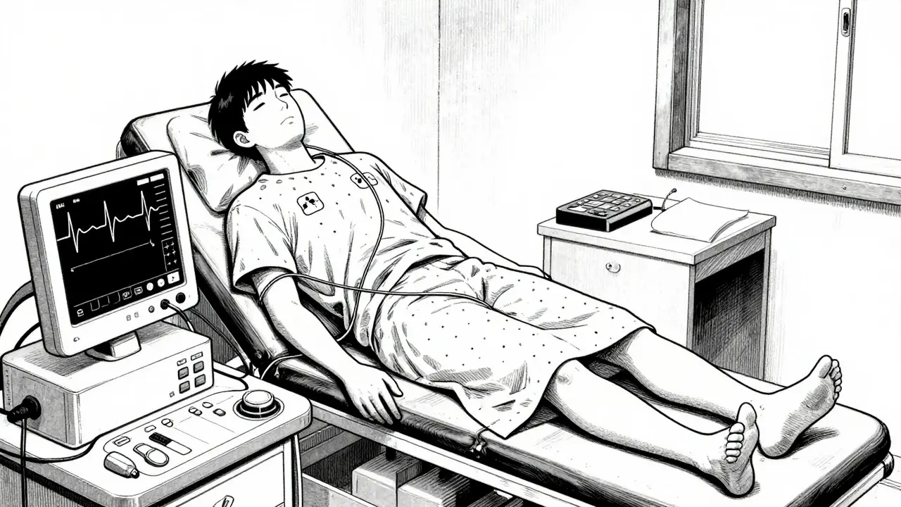
An Electrocardiogram measures the heart's electrical signals using small sensors called electrodes attached to your skin. Developed back in 1903 by Willem Einthoven, who won a Nobel Prize for this work, the technique hasn't changed much in concept, though the digital recording is vastly improved. During a standard session, sticky pads go on your chest, arms, and legs. The machine translates the tiny voltage changes into squiggly lines on a screen or paper. This takes only three to five minutes, yet it provides a snapshot of your rhythm, rate, and conduction patterns.
This isn't just for people currently having a heart attack. Doctors use these readings to spot irregularities like atrial fibrillation, past heart damage, or thickening of the heart walls. However, a resting ECG has limits. If your heart functions normally while you sit quietly, a standard reading might miss problems that only appear when you demand more energy from your muscles.
Pushing Limits with Stress Testing
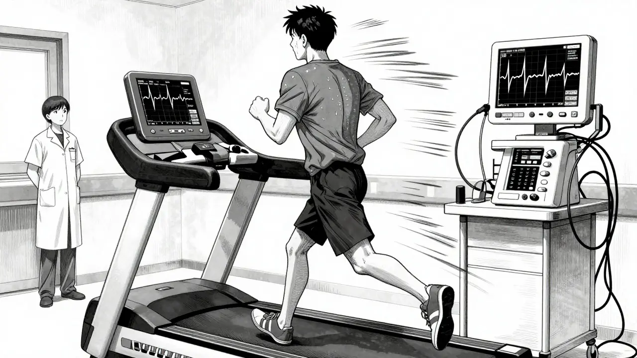
That is where the exercise stress test enters the picture. While a resting ECG looks at your engine idling, a stress test sees how it performs at high RPMs. Formally known as an exercise electrocardiogram, this procedure evaluates how your heart reacts to physical exertion. The logic is simple: healthy arteries dilate and supply blood easily, but narrowed vessels struggle under pressure, causing oxygen shortages.
Most clinics use the Bruce Protocol. This is a standard treadmill method that starts slow and gets harder every few minutes. You begin walking at 1.7 miles per hour with a slight incline. Every three minutes, both speed and grade increase until you reach your target heart rate. That target is usually calculated as 220 minus your age, aiming for about 85% of your maximum capacity.
Why the increasing difficulty? Because significant blockages might stay hidden during light activity. Only when the heart works hard does it signal distress, usually seen as dips in the electrical waves or drops in blood pressure. According to major cardiology standards, over 11 million of these tests happen annually in the U.S. alone, proving their value in spotting issues like coronary artery disease that a static scan misses.
| Type | Radiation Exposure | Sensitivity for CAD | Ideal Patient Profile |
|---|---|---|---|
| Exercise ECG | None | ~68% | Active, able to walk/run |
| Stress Echocardiogram | None | ~82% | Moderate ability, women, intermediate risk |
| Nuclear Stress Test | 9-12 mSv | ~89% | Able to exercise or pharmacological option needed |
The choice between these methods depends largely on your lifestyle and physical condition. An exercise ECG is the go-to if you can walk briskly without stopping. However, if arthritis, asthma, or mobility issues prevent you from hitting that target heart rate on a treadmill, doctors switch tactics. Instead of movement, they use medication to simulate the stress. Agents like adenosine or dobutamine are pumped into your vein, artificially stressing the heart muscle so the imaging can capture any blood flow restrictions.
Decoding the Results: Numbers and Waves
Once the test is finished, the real work begins for the cardiologist. You might see terms like "ST-segment depression" on the report. In plain English, this suggests the heart muscle isn't getting enough oxygen during peak effort. The duration you lasted on the treadmill is also a huge indicator. Research shows that for every additional minute of exercise you complete, your risk of a future cardiac event drops by roughly 12%. Functional capacity is a vital sign.
Accuracy varies depending on the technology used. A standard stress ECG catches about two-thirds of cases where coronary artery disease exists. It's good, but it can miss things, especially in women whose physiology sometimes produces different signals. This is why combining the stress component with imaging helps. Stress echocardiography adds ultrasound visuals to the mix, raising the accuracy significantly without using radiation. Meanwhile, nuclear testing gives the highest sensitivity-detecting nearly 90% of blockages-but comes with a price tag of radiation exposure equivalent to a few years of background sunlight.
Determining which test is right involves looking at your pretest probability. If a doctor thinks you are unlikely to have heart issues based on your symptoms, they might not order a stress test at all. Conversely, if you have moderate symptoms, guidelines suggest starting with functional testing rather than jumping straight to expensive CT scans or invasive angiograms. The goal is to rule out serious danger efficiently without unnecessary procedures.
What the Experience Feels Like
Patient stories paint a clearer picture than technical manuals. Many people are surprised by the simplicity of the setup. You wear a gown, get hooked up to wires, and step onto the treadmill. Some describe it as surprisingly easy, lasting ten to twelve minutes before exhaustion or target heart rate ends the run. Others feel nervous watching the monitor flash numbers. It is normal to feel anxious, but the technicians are trained to spot warning signs instantly.
If you undergo the chemical version, the experience is different. Medications like adenosine flood the system with sensations that mimic anxiety: shortness of breath, chest pressure, and flushing. These feelings peak within minutes and pass quickly once the drug wears off. Knowing this beforehand prevents panic. About 15% of patients report unpleasant side effects from the chemicals, but the safety profile remains robust when monitored correctly.
Preparing for the Appointment
Getting ready for the test matters just as much as the test itself. You want clean data, not interference. Avoid caffeine entirely for 24 hours before your appointment. Caffeine can alter your heart rate response and mess with the drugs used in pharmacological stress tests. Wear comfortable clothes that allow free movement, even though you'll likely change into a gown. Bring a list of your medications, as some affect how your heart reacts to stress.
Don't eat a heavy meal beforehand. A full stomach makes exercising harder and could lead to nausea during the strain. Arrive relaxed. The staff knows what to look for, and the environment is controlled. Once you leave, you can usually resume your normal routine immediately, assuming no complications arose during the procedure.
Limitations and False Signals
No test is perfect, and understanding the flaws helps manage expectations. Resting ECGs can produce false positives due to anxiety or minor electrical variations unrelated to blockages. Conversely, they can miss microvascular dysfunction, where tiny blood vessels fail to function even if the big arteries look open. This is particularly tricky in premenopausal women, whose symptoms might be triggered by smaller vessel issues that standard imaging misses.
Technology is evolving to fix this. Artificial intelligence is being integrated into cardiology software now. Recent studies from 2023 suggest AI algorithms can boost interpretation accuracy by over 20%, catching subtle patterns human eyes overlook. Portable devices are also emerging, allowing testing outside the hospital room, though standard treadmill setups remain the gold standard for diagnostic reliability.
Your Next Steps After Diagnosis
If the test shows clear blockages, the doctor might recommend further steps like a coronary angiogram, which is a more invasive procedure involving dye and catheters. If the results are inconclusive, you might get a repeat test or a different modality, like a CT angiogram. If everything looks normal despite your symptoms, the doctor might look at non-cardiac causes for your chest pain, such as musculoskeletal strain or gastrointestinal issues.
Keep your records. Tracking how long you lasted on the treadmill year over year gives you a personal baseline for your fitness and heart health. Regular check-ups are not just about finding disease early; they are about maintaining the capacity to keep pumping blood effectively throughout your life.
Is an ECG painful?
No, a resting ECG is completely painless. It only requires placing stickers on your skin to read electrical signals. You remain still, and the process takes less than five minutes.
Can I drink coffee before a stress test?
You should avoid caffeine for at least 24 hours prior to testing. Caffeine affects heart rate and can interfere with the stress medications or obscure the results.
What happens if I have low fitness levels?
If you cannot exercise on a treadmill due to joint issues or poor fitness, doctors use a chemical stress test. A medication simulates the effect of exercise so they can still image your heart's blood flow.
Does a stress test involve radiation?
A standard exercise ECG does not use radiation. However, a nuclear stress test involves a radioactive tracer. Stress echocardiograms use ultrasound sound waves and are also radiation-free.
When do I get the results?
Many facilities provide preliminary findings right after the test. Final reports are usually reviewed by a cardiologist and sent to your primary doctor within 24 to 48 hours.